APD based technology with Depth-of-Interaction capability
What is the benefit of Time-of-Flight?
Our Avalanche Photodiode (APD) based technology for PET was developed by a R&D Consortium including PETsys S.A., with a focus on research and development. The R&D Consortium licensed this technology to PETsys S.A.
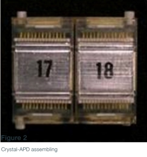
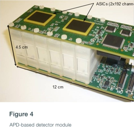
The APD-based photon detectors use matrices of LYSO scintillator crystals coupled to APD arrays. The APD signals are readout and sampled by a dedicated ASIC with 192 channels.
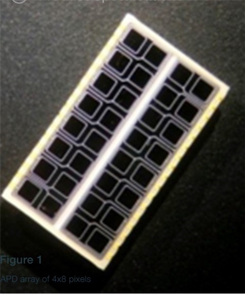
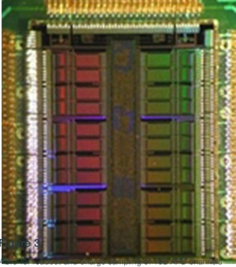
Several LYSO APD-dots and the corresponding channel activation pattern in mini-module = detector modules. Our APD-based modules contain two mini detector modules, each with 192 channels.
One of the technologies we apply is the capability of determining the depth-of-interaction (DOI) of the photons within the crystal. This technique, based on the separation of scintillation light into the APD pixels for different interaction depths, allows elimination of parallax errors in reconstructed space positions of the events. It is not easy to apply this crystal coating technique on diced crystal matrices.
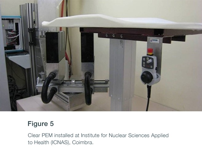
PETsys has developed two PET prototypes based on APD-based detector modules which target breast cancer applications (Clear PEM System).
These systems comprise 12288 APDs and 6144 scintillator crystals, organized in two plates. The front-end electronics. DOI of Trues is included in CASTOR to Customize PET - Fig. 5
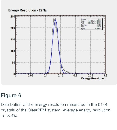
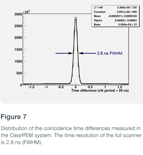
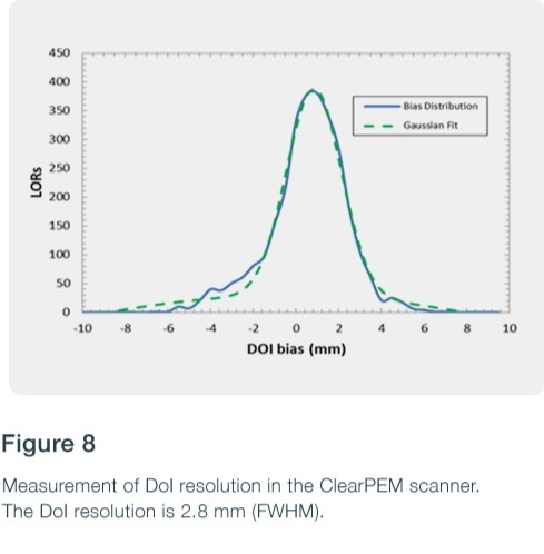
The main performance characteristics of the Clear PEM scanner include an energy resolution (Er) of 15.9% and a DOI precision of 2.2 mm, assuring view resolution of ~1.3 mm (FWHM).
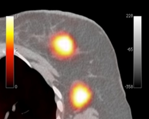
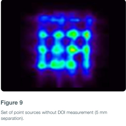
Clear PEM image results without shown above calculation of depth-of-interaction (left) vs. with depth resolution of 5-10 mm (right). Images of the NEMA breast phantom on a derenzo insert. Compare with 18F-FDG image resolution of ~1.3 mm (FWHM).


Due to its very good spatial resolution and accessibility of the Field-of-View, the Clear-PEM system can also be used for small animal imaging.